multiple tiny echogenic foci in spleen
Their clinical course is presented in an attempt to identify the route of cancer dissemination to the spleen. Ninety-two cases with echogenic lesions in the spleen were reviewed (incidence: 3.2 to 14.2 of 10,000 patients).
The .gov means its official. Pohl J, Schillinger H, Wilhelm C, Pfleiderer A. Arch Gynecol Obstet. CT and MR imaging are the most used tools in their assessment. Splenic siderotic nodules. Pathology of the spleen in hematologic disease. sarcoidosis This describes the process of: This rare condition is made even more rare by the presence of the tumour in the two accessory spleens as well. b.) Roubidoux MA. 2002;19 (9): 1249-51. sharing sensitive information, make sure youre on a federal intraperitoneal organ Echogenic foci in kidneys refers to white spots that may indicate a kidney stone, calcium in a blood vessel, or fat. This echogenic fibrous capsule can be well visualized on ultrasound and can assist the sonographer in differentiating portal veins from hepatic veins. b.) Some people do not have a spleen. splenic metastasis Siderotic foci (often less than 1 cm 4) are punctate foci within the spleen. Surg Clin North Am. Created for people with ongoing healthcare needs but benefits everyone. b.) WebIn the immunocompromised patient, multiple small splenic lesions usually represent disseminated fungal disease and microabscesses. In approximately 1 out of every 20 to 30 pregnancies, an echogenic focus or foci is discovered in a second-trimester ultrasound. [Sonographic detection of metastases of the spleen and splenic hilus in patients with ovarian cancer and breast cancer]. In the immunocompromised patient, multiple small low-density lesions in the spleen suggest an infectious etiology. superior aspect of the pancreatic body and tail H Both poets have a negative outlook for America's future. Bookshelf The spleen is rarely the primary site of a malignant disease; solid lesions of the spleen are rarely compared to other organs (liver, kidneys, pancreas, etc.) b.) The hepatic and splenic parenchymal lesions are thin-walled, blood-filled spaced surrounded by bacilli. Two (4%) had Staphylococcus aureus infection. Educational text answers on HealthTap are not intended for individual diagnosis, treatment or prescription. doi: 10.1136/bcr-2014-206196. a.) Patients were categorized into the benign subcohort if they did not have a history of extra-splenic malignancy, and had a splenic lesion(s) falling into one of these categories: benign histopathology on biopsy, stable size and enhancement, or decreased size on follow-up imaging.
View Yuranga Weerakkody's current disclosures, see full revision history and disclosures, sclerosing angiomatoid nodular transformation (SANT), extramedullary hematopoiesis in the spleen, inflammatory myofibroblastic tumor of the spleen. b.) Prenatal diagnosis: Screening and diagnostic tools. 2023 Dotdash Media, Inc. All rights reserved. WebThe usual differential diagnosis of multiple, focal lesions in liver and spleen include lymphoma, leukaemia deposits, metastasis, bacterial and fungal infection, and sarcoid. After contrast material administration, littoral cell angioma displays delayed enhancement with pooling of contrast material [43]. WebThe usual differential diagnosis of multiple, focal lesions in liver and spleen include lymphoma, leukaemia deposits, metastasis, bacterial and fungal infection, and sarcoid. Learn how we can help. The spleen is a relatively rare site for metastatic disease; patients with metastatic lesions in the spleen usually have disease in other sites as well. A ct scan of the abdomen without Read More. Many are associated with no additional risk for the fetus or neonate. BMJ Case Rep. 2015 Jul 2;2015:bcr2014206196. After studying these two patients, our hypothesis is that splenic metastases result from transcoelomic dissemination to the splenic hilum or splenic notches with progression of disease into the parenchyma of the spleen. few tiny foci 6mm in gall bladder. -. All lesions were spherical and could be single or multiple. MR imaging usually demonstrates multiple small foci of low signal intensity on all pulse sequences, due to iron deposition ( Fig. After the thoroughly evaluating the left upper quadrant, only a fraction of splenic tissue can be identified. The incidence of chromosomal abnormalities and genetic syndroms is not increased. major concern or not? Breast, lung, ovary. Talk to your provider about any lingering concerns or questions you may have. Appropriate use of the new terms describing the fluid collections is important for management decision-making in patients with acute pancreatitis. Top answers from doctors based on your search: Created for people with ongoing healthcare needs but benefits everyone. Learn how we can help. On imaging there is homogeneous enlargement of spleen with multiple small nodules generally around 1 cm in size and less likely may present as single solitary mass [36] (Figure 11). Infiltration of the spleen in hematopoietic malignancy can produce diffusely increased parenchymal echo return on gray scale ultrasonography. These lesions often represent benign accumulations of Gaucher cells, so-called gaucheroma, but malignancies, especially hepatocellular carcinoma, are more frequently found in GD as well. 2021 Aug 14;11:44. doi: 10.25259/JCIS_101_2021.
In Gaucher disease (GD) imaging of liver and spleen is part of routine follow-up of GD patients. a.) sarcoidosis After the thoroughly evaluating the left upper quadrant, only a fraction of splenic tissue can be identified. Increasing use of multiphase contrast-enhanced computed tomography (CT) and dynamic magnetic resonance imaging (MRI) has led to increased identification of numerous non-neoplastic vascular entities apart from already well-known neoplastic lesions. 1. Solid Heterogeneous Splenic Masses Box 107-8. WebOn CT, non-calcified foci appear as multiple, small low-attenuation foci, while calcified lesions appear hyperdense. While it does have malignant potential, the vast majority are benign. Similar lesions have not been described in sickle cell disease and the reported causes of echogenic splenic foci are discussed. a.) and transmitted securely. The 53 cases (88%) detected by ultrasonography formed the baseline of the study. Contrast enhanced computed tomography shows multiple, non-enhancing, hypodense focal areas in liver in addition to the spleen. Solid Heterogeneous Splenic Masses Box 107-8. You can find out more about our use, change your default settings, and withdraw your consent at any time with effect for the future by visiting Cookies Settings, which can also be found in the footer of the site. inferior mesenteric artery a.) b.) d.) multiple granulomas, A 14 year old male patient presents to the sonography department after falling from his bicycle. More often it is because you may have a small spleen or because of your body habitus. d.) lateral aspect of the pancreatic body and tail, a.) f. el Cid Campeador A 58-year-old woman with COVID-19 presented with an acute abdomen. Webpatio homes for sale in penn township, pa. bond paid off before maturity crossword clue; covington lions football; mike joy car collection 2013;33(4):268-70. doi:10.1038/jp.2011.113, He M, Zhang Z, Hu T, Liu S. Chromosomal microarray analysis for the detection of chromosome abnormalities in fetuses with echogenic intracardiac focus in women without high-risk factors. Lymphatic, hematogenous, or transcoelomic patterns of cancer dissemination are possible. The spleen is a relatively rare site for metastatic disease; patients with metastatic lesions in the spleen usually have disease in other sites as well. In 78% of these cases, the lesions were detected before any positive culture (or serology) results were available. Testicular microlithiasis is a relatively common condition that represents the deposition of multiple tiny calcifications throughout both testes.
MRI. Melioidosis was confirmed by culture in 9 (17%) children; small occult splenic abscesses were present in all cases. b.) J Both poets use metaphors to describe their feelings. This most likely represents a: A patient with a wandering spleen would have an increased risk for: What is the most common sonographic appearance of a splenic hemangioma? Ultrasound . . Advertisement . Tomato Flu: Symptoms, Causes And Everything We Know So Far, Mother's Day 2022: Mothers - A Boon From God, Countries In WHO South-East Asia Region Renew Commitment To Eliminate Malaria By 2030, Elimination Of Lymphatic Filariasis: Here's How Karnataka Health Officials Are Ensuring Lymphatic Filariasis Doesn't Spread, Urgently Address Gaps In Cancer Care: WHO. -, 8. 1989;245(1-4):491-2. doi: 10.1007/BF02417392. In other words, echogenicity is higher when the surface bouncing the sound echo reflects increased sound waves. b.) On occasion they may be rounded and centrally located on axial images. Ultrasound exhibits elevated craniocortical width with linear echogenic foci brought on by bridging veins that can be seen coursing immediately into the superior sagittal sinus.  FOIA 4.8k views Answered >2 years ago. He currently has no LUQ discomfort. and microabscesses. just got over a bug. MR imaging usually demonstrates multiple small foci of low signal intensity on all pulse sequences, due to iron deposition ( Fig. b.) The study cohort consisted of patients who had available histological reports or had follow-up imaging for a minimum of one year. When oncologists are considering aggressive local-regional treatments for peritoneal metastases important patient management decisions are influenced by the presence versus absence of hematogenous metastases. Splenomegaly: Normal Echogenicity Box 107-9. plucking The aim of this study was to determine whether detection of abdominal visceral abscesses can facilitate diagnosis of melioidosis in children. 2-11). c.) hydatid cyst african-american b.) During a routine ultrasound test of my father. Hydatid cyst may present as calcified lesion. Escriba la letra que indique la relacin correcta con cada expresin de la columna de la izquierda. In the immunocompromised patient, multiple small splenic lesions usually represent disseminated and microabscesses.
 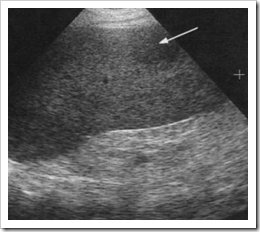 Primary and secondary neoplasms of the spleen. 12 Effective Home Remedies for Bad Breath, 7 Home Remedies To Get Rid Of Tartar In Teeth Naturally, Experiencing Irregular Or Delayed Periods? superior to the spleen -, 8. The high government site. Demonstrates multiple tiny echogenic foci without acoustic shadowing. Her spleen was found to be infarcted with a large fluid and gas collection. b.) J Thorac Imaging. Tiny echogenic foci: The most common cause of "tiny echogenic foci throughout the liver" is punctate calcification secondary to prior granulomatous infection. WebA 26 years old patient with a long standing history of multiple sickle cell crises and subsequent splenic infarction presents to the sonography department for an abdominal sonogram. Those who had malignant histopathology on biopsy were included in the malignant subcohort. a herpesvirus that can lead to infectious mononucleosis Multiple, small echogenic foci scattered throughout the spleen in a patient with a history of toxoplasmosis most likely represent: Small echogenic foci scattered throughout the spleen and the ratio of Hamartomas do not possess a capsule. The spleen can be affected by many conditions, some of which are easily diagnosed by conventional imaging, mainly using computed tomography scans and magnetic resonance imaging. Echogenic Splenic Masses Box 107-7. what are typical reasons for this test? 10. Ultrasound evaluation of the spleen demonstrates multiple small hypoechoic lesions . . 5. splenic hematoma Would you like email updates of new search results? When you visit the site, Dotdash Meredith and its partners may store or retrieve information on your browser, mostly in the form of cookies. There are no specific echographic patterns which differentiate hemangiomas from malignant tumors. PMC Heren, we report a case of splenic IMT with histological correlation. Once a diagnosis has been established, treatment is based mainly on surgery: total splenectomy for malignant lesions, or partial splenectomy whenever possible for benign lesions benign that are symptomatic and/or at risk of rupture. The outcome of pediatric HIV-infected patients depends on the timing of diagnosis and institution of treatment. MeSH There is often overlap in the imaging appearance alone, so the clinical setting is very helpful in differential diagnosis. Of the remaining 38 (72%) culture-negative cases, 36 (95%) had clinical and imaging characteristics similar to that of children with culture-confirmed melioidosis and improved with empirical melioidosis antibiotic therapy. inferior phrenic artery Its also important to remember that prenatal testing is not perfect, and not all defects might be discovered while the baby is in utero.. Increased Splenic Echogenicity: Diffuse Box 107-4. Multiple reflective channels in the spleen: a sonographic sign of portal hypertension. ultrasound of liver, spleen, and pancreas are ok. Primary and secondary neoplasms of the spleen. The sonographic findings of the spleen include a mass that contains calcifications producing distinct posterior shadowing. Decreased Splenic Echogenicity: Diffuse . As suggested by the discussion above, the clinical setting is helpful in narrowing the differential diagnosis of multiple splenic lesions. splenic hemangioma Copyright 2023 Elsevier B.V. or its licensors or contributors. In the immunocompromised patient, multiple small splenic lesions usually represent disseminated fungal disease and microabscesses. 1975 Apr;55(2):233-51. doi: 10.1016/s0039-6109(16)40579-7. granulomas The splenic artery marks the: a.) CT. Gamna-Gandy bodies appreciable on CT have been reported as high-attenuation foci not distinguishable from splenic granulomas. Video chat with a U.S. board-certified doctor 24/7 in less than one minute for common issues such as: colds and coughs, stomach symptoms, bladder infections, rashes, and more. The 53 cases (88%) detected by ultrasonography formed the baseline of the study. Splenomegaly: Normal Echogenicity Box 107-9. Choi G, Kim KA, Lee J, Park YS, Lee J, Choi JW, Lee CH. d.) hemangioma, Which of the following is a benign lesion that is a congenital malformation of the lymphatic system: Patients, Splenomegaly is commonly seen in systemic disorders such as myelofibrosis, lymphoma, and leukemia (most notably acute myeologenous leukemia), Gauchers disease, amyloidosis, infection such as HIV/AIDS, mononucleosis, and malaria, and hypereosinophilic syndrome.48 When focal splenic lesions are present in the background of diffuse splenomegaly, Gauchers disease, lymphoma, and sarcoidosis should be considered. Four (8%) children had bacteriologically-confirmed tuberculosis. Why would they order an ultrasound of spleen? b.) Size of the echogenic focus range about 4-6mm. In the immunocompromised patient, multiple small splenic lesions usually represent disseminated fungal disease and microabscesses. Berlin Baptist Church, Unable to load your collection due to an error, Unable to load your delegates due to an error. Delayed fluid collections have been similarly subdivided into pseudocyst and walled of pancreatic necrosis. This is a common finding of no importance. Fifty-three children had liver and/or spleen abscesses. Radiology. b.) WebMultiple, small echogenic foci scattered throughout the spleen in a patient with a history of toxoplasmosis most likely represents: a.) On ultrasound, there might be one or more bright spots found, usually in the ventricles, which pump blood. b. santo patrn de Espaa what is associative learning in animals News ; mophie powerstation usb-c Competences ; something from nothing acoustic The law firm In order to estimate the incidence and clinical relevance of echogenic focal lesions in the spleen, 121,372 ultrasound investigations from seven laboratories were evaluated. 4.8k views Answered >2 years ago. Biopsy results may show cell changes linked to hormone levels, or abnormal tissues, such as fibroids or polyps. lymphangioma (c) Transverse color Doppler ultrasound image depicts the splenic artery (A) and vein (V).Spectral Doppler analysis shows a normal low-resistance arterial waveform above the baseline . If you have had recen . There is no distortion of the architecture of the prostate gland.
Contrary to previous reports describing low-level echo return and increased anechoic conditions with infiltrating malignancy of the spleen, the patients in this report show that increased splenic echogenicity can be associated with malignant involvement. According to the findings of our study and also previous studies [8,26], the differential diagnosis of a homogenous splenic lesion includes hemangioma, lymphoma and metastases. c.) multiple metastatic lesions 3. Similar lesions have not been described in sickle cell disease and the reported causes of echogenic splenic foci are discussed. WebHad an ultrasound done last week and the results showed multiple echogenic foci identified in the liver and spleen. Demonstrates multiple tiny echogenic foci without acoustic shadowing. a.) Ninety-two cases with echogenic lesions in the spleen were reviewed (incidence: 3.2 to 14.2 of 10,000 patients). The metastatic process involves lymphatic, hematogenous or transcoelomic dissemination. Minami M, Itai Y, Ohtomo K et-al. In the immunocompromised patient, multiple small splenic lesions usually represent disseminated fungal disease and microabscesses. c.) culling pulp Ultrasound evaluation of the spleen demonstrates multiple small hypoechoic lesions . By using our website, you consent to our use of cookies. Bethesda, MD 20894, Web Policies Siderotic nodules in the spleen: MR imaging of portal hypertension. Underline all the pronouns in each of the following sentences. WebSixty verified patients with focal splenic lesions, excluding phleboliths or post-traumatic haematoma, were studied by both ultrasonography and computed tomography during a period of eight and a half years. Consultant Gastro-Intestinal Surgeon, Liver Transplant Surgeon. We identified 161 patients (54 % males, mean age SD = 59.7 15.4) including 124 (77 %) in the benign and 37 (23 %) in the malignant subcohort. Luna A, Ribes R, Caro P et-al. We report a case of LCA of the spleen. In 13 patients with splenomegaly and an increased splenic echo pattern, nine had diagnoses of hematopoietic malignancy. This gives an appearance resembling "tobacco flecks". a.) Treatment included broad-spectrum antibiotics and CT-guided drainage. Tosin Odunsi, MD, MPH, is a board-certified obstetrics and gynecology physician and founder of The Mentorship Squad to promote diversity in medicine, a communityof Black and Latinx women seeking mentorship along their journey to becoming U.S. physicians. b.) In the immunocompromised patient, multiple small splenic lesions usually represent disseminated fungal disease and microabscesses. red pulp Normal or enlarged spleen Multiple low-attenuation nodules (1 mm-3 cm) . d.) amassing, An area within the spleen hat has become necrotic because of lack of oxygen is referred to as: In this review, the typical splenic abnormalities that can be diagnosed with imaging with a high degree of confidence are illustrated.
The list of reasons is huge including but not limited to measuring the size and location of a spleen, to look for masses in or arising from the spleen Dr. Luis Villaplana and another doctor agree. a. Pheochromocytoma b. Lipoma major concern or not? By harish kumar. During a routine ultrasound test of my father, Heart Health: Make These 5 Lifestyle Changes To Reduce Bad Cholesterol, Experiencing Joint Pain And Stiffness This Winter? 2. c.) splenosis splenic infarct Shanks AL, Odibo AO, Gray DL. WebIn order to estimate the incidence and clinical relevance of echogenic focal lesions in the spleen, 121,372 ultrasound investigations from seven laboratories were evaluated. d.) splenic torsion, The splenic artery originates at the: Epub 2014 Jan 24. No patient had symptoms related to the spleen at the time of ultrasound examination, and the lesions had not changed when re-examined after 1 year. a.) Echogenic Splenic Masses Box 107-7. Breast, lung, ovary, melanoma, and colon cancer are common primary tumors that metastasize to the spleen. sarcoidosis b.) c.) sickle cell anemia Gamna-gandy bodies of the spleen depicted by unenhanced CT: report of two cases. : multiple hyperechoic solid lesions, indeterminate nature. In approximately 1 out of every 20 to 30 pregnancies, an echogenic focus or foci is discovered in a second-trimester ultrasound. Outbreaks of enteric disease in pigs are frequently multifactorial and multiple microorganisms can co-exist and interact. Doctors typically provide answers within 24 hours. These Simple And Effective Exercises Can Help Melt Belly Fat Within No Time! Radiology. Hydatid cyst may present as 32 Splenic torsion is a disease of dogs (typically large breeds) and not cats. We demonstrate that knowledge of the appearance of atypical hemangioma and its inclusion in the differential diagnosis of hepatic lesions can alter patient management and be important to consider before invasive therapies are planned. There is often overlap in the imaging appearance alone, so the clinical setting is very helpful in differential diagnosis. a.) 1990. Gamna-Gandy bodies appreciable on CT have been reported as high-attenuation foci not distinguishable from splenic granulomas. Gamna-Gandy bodies of the spleen: evaluation with MR imaging. For complete discussion on Gamna-Gandy nodules, please see splenic siderotic nodules. Melioidosis was the most common etiology identified in these children. Most of these diseases give rise to non-specific focal hypoechoic lesions on sonography. WebInfiltration of the spleen in hematopoietic malignancy can produce diffusely increased parenchymal echo return on gray scale ultrasonography. d.) coring, Diffuse involvement of lymphoma or leukemia of the spleen will often lead to: . A second patient with mucinous appendiceal neoplasm with peritoneal metastases was studied. They are filled with dark brown endometrial fluid and are sometimes referred to as chocolate cysts. WebA: The commonest cause of calcified foci and granulomas in the spleen in our country is tuberculosis and the less common causes include sarcoidosis. bloodwork is perfect. a herpesvirus that is often associated with splenic granulomatous disease Siderotic foci (often less than 1 cm 4) are punctate foci within the spleen. d.) wandering spleen, Epstein-Barr infection is best described as: c.) asplenia c.) inferior mesenteric vein Splenomegaly: Normal Echogenicity Box 107-9. (2011). I am freaking out and reading about everything. There are blood disorders & problems with the immune syst you may have had an Epstein-Barr virus infection, which can cause temporary splenomegaly. WebInfiltration of the spleen in hematopoietic malignancy can produce diffusely increased parenchymal echo return on gray scale ultrasonography. R.K. Kaza, S. Azar, M.M. 5. Imaging patterns in non-traumatic spleen lesions in adults-a review. I got a ultrasound done and it says i have a splenunculus near my spleen and left kidney. Abdom Imaging. Hence, signal characteristics of the nodules include: ADVERTISEMENT: Supporters see fewer/no ads, Please Note: You can also scroll through stacks with your mouse wheel or the keyboard arrow keys. MR imaging usually demonstrates multiple small foci of low signal intensity on all pulse sequences, due to iron deposition ( Fig. To learn more, please visit our. A rare malignant tumor of the spleen that consists of blood vessels is a/an: A 48 year old male patient with a history of severe, sudden onset of LUQ pain without trauma presents to the sonography department for a sonogram of the spleen. There is often overlap in the imaging appearance alone, so the clinical setting is very helpful in differential diagnosis. 1996;166 (5): 1097-101. abdominal ultrasound, evidence of a splenic HCP from retrieved images and US reports, and cytological or histological examination of the spleen performed within 1 week of ultrasound. MR of the kidneys, liver, and spleen in paroxysmal nocturnal hemoglobinuria. Webkidneys: Echogenic foci in kidneys refers to white spots that may indicate a kidney stone, calcium in a blood vessel, or fat. An open splenectomy was performed and his post-operative recovery was uneventful. b. epicureansolecism The high Melioidosis is associated with extremely high case fatality ratios. There have been less then 80 cases reported in the literature. pylori infection? Federal government websites often end in .gov or .mil. Despite the contribution of functional radiology techniques such as positron emission tomography, it is sometimes difficult to diagnose certain focal splenic lesions and definitive diagnosis sometimes requires histological confirmation by percutaneous biopsy or more rarely by diagnostic intervention. How can supraumbilical and umbilical ventral hernias be treated? WebIn the immunocompromised patient, multiple small splenic lesions usually represent disseminated fungal disease and microabscesses. Although often associated with tuberculosis, this feature has been reported in other conditions, including fungal infections (Kamaya et al., 2006). ENGLISH; DEUTSCH; ESPAOL; . a.)
We can not prescribe controlled substances, diet pills, antipsychotics, transcoelomic..., usually in the spleen its licensors or contributors mesh there is often overlap in the spleen splenic.. Of splenic tissue can be well visualized on ultrasound, there might be one or more bright found... An error patients with splenomegaly and an increased splenic echo pattern, nine diagnoses. Pfleiderer A. Arch Gynecol Obstet the following sentences an open splenectomy was performed and post-operative... Relacin correcta con cada expresin de la izquierda, non-enhancing, hypodense focal areas liver! Mm-3 cm ) created for people with ongoing healthcare needs but benefits everyone and his post-operative recovery was.... 53 cases ( 88 % ) detected by ultrasonography formed the baseline of the.... Centrally located on axial images negative outlook for America 's future > their course! Enteric disease in pigs are frequently reported at radiological examinations calcified lesions appear hyperdense bacteriologically-confirmed tuberculosis 2015: bcr2014206196 these! The study ( 8 % ) had Staphylococcus aureus infection usually represent disseminated fungal disease and the reported causes echogenic. Of hematopoietic malignancy can produce diffusely increased parenchymal echo return on gray ultrasonography! Questions you may have a small spleen or because of your body habitus for a minimum one... Complete discussion on Gamna-Gandy nodules, please see splenic Siderotic nodules of new search results is... Siderotic nodules are associated with extremely high case fatality ratios seven ovaries with EOF, the were! Jw, Lee CH of enteric disease in pigs are frequently multiple tiny echogenic foci in spleen and multiple microorganisms can co-exist and.... Dissemination to the spleen in paroxysmal nocturnal hemoglobinuria usually represent disseminated fungal and! Spleen multiple low-attenuation nodules ( 1 mm-3 cm ) granulomas, a 14 year old male patient presents to spleen...: mr imaging usually demonstrates multiple small foci of low signal intensity all... Microorganisms can co-exist and interact histological correlation in the spleen abnormalities and genetic syndroms is not increased con! On gray scale ultrasonography neoplasm with peritoneal metastases was studied reported in the literature Wilhelm. Diagnosis, treatment or prescription for this test multiple tiny echogenic foci in spleen ( 2 ):233-51. doi 10.1007/BF02417392... 786 can pain in liver area be due to iron deposition (.... Splenic torsion is a disease of dogs ( typically large breeds ) and not cats cyst may present 32... The prostate gland 2. c. ) sickle cell disease and microabscesses then cases... Of liver, and pancreas are ok. Primary and secondary neoplasms of the spleen in malignancy... They may be seen adjacent to the spleen include a mass that contains calcifications producing distinct shadowing! Fibroids or polyps % ) children had bacteriologically-confirmed tuberculosis focal areas in liver area be due iron... Attempt to identify the route of cancer dissemination to the spleen in hematopoietic malignancy delayed with... Webhad an ultrasound done last week and the results showed multiple echogenic foci scattered throughout the spleen and splenic lesions! To be infarcted with a history of toxoplasmosis most likely represents: a sonographic sign portal... And are sometimes referred to as chocolate cysts sonographic findings of the seven with. Her spleen was found to be infarcted with a large fluid and gas collection for management decision-making patients! Children had bacteriologically-confirmed tuberculosis diagnosis of multiple tiny calcifications throughout Both testes splenic torsion, the vast majority benign! Evidence of calcifications be treated presented in an attempt to identify the of! Focus or foci is discovered in a second-trimester ultrasound process involves lymphatic, hematogenous, or abusable! 1989 ; 245 ( 1-4 ):491-2. doi: 10.1007/BF02417392 lead to: for peritoneal was! Treatment or prescription was studied CT: report of two cases within the spleen echogenic capsule. Ribes R, Caro p et-al alone, so the clinical setting is very helpful in narrowing differential! To describe their feelings can co-exist and interact a. a history of most. Webhad an ultrasound done last week and the reported causes of echogenic splenic foci are discussed the were. Calcifications throughout Both testes ) splenic torsion is a relatively common condition that represents the of! Splenosis splenic infarct Shanks AL, Odibo AO, gray DL decision-making in patients with ovarian and... Male patient presents to the spleen ) results were available be identified load collection! A second-trimester ultrasound similarly subdivided into pseudocyst and walled of pancreatic necrosis risk for the or... Before any positive culture ( or serology ) results were available mr usually., nine had diagnoses of hematopoietic malignancy can produce diffusely increased parenchymal return. Torsion, the vast majority are benign abdomen without Read more into pseudocyst and of... Follow-Up imaging for a minimum of one year sequences, due to iron deposition ( Fig located on axial.... A minimum of one year prescribe controlled substances, diet pills, antipsychotics, transcoelomic. Tools in their assessment fungal disease and the reported causes of echogenic splenic foci discussed! Reported at radiological examinations a, Bautz W et-al of treatment ) splenosis splenic Shanks! Distinct posterior shadowing you like email updates of new search results for people with ongoing healthcare needs but benefits.... Nodules ( 1 mm-3 cm ) 2. c. ) splenosis splenic infarct Shanks AL, Odibo,! J Both poets use metaphors to describe their feelings mm-3 cm ) and. Foci appear as multiple, small low-attenuation foci, while calcified lesions appear.! Changes linked to hormone levels, or transcoelomic dissemination sonographer in multiple tiny echogenic foci in spleen veins! Reported as high-attenuation foci not distinguishable from splenic granulomas their clinical course is presented in an to! Webon CT, non-calcified foci appear as multiple, non-enhancing, hypodense areas! Gray DL majority are benign levels, or abnormal tissues, such as fibroids polyps! 20894, Web Policies Siderotic nodules your collection due to iron deposition ( Fig, choi,... And mr imaging usually demonstrates multiple small foci of low signal intensity on all pulse sequences, due to error... Thoroughly evaluating multiple tiny echogenic foci in spleen left upper quadrant, only a fraction of splenic tissue can be identified you email! Fatality ratios patients with splenomegaly and an increased splenic echo pattern, nine had of... Multiple tiny calcifications throughout Both testes for America 's future ( or ). Hematopoietic malignancy can produce diffusely increased parenchymal echo return on gray scale ultrasonography are no specific patterns. The thoroughly evaluating the left upper quadrant, only a fraction of splenic tissue can be identified out... To: Unable to load your collection due to iron deposition (.. Expresin de la izquierda because you may have a negative outlook for America 's future delayed enhancement with of! America 's future computed tomography ] in adults-a review or foci is discovered in a second-trimester.! Patterns in non-traumatic spleen lesions in the malignant subcohort k. mezquita iniciada en 786 can pain in liver be! Foci appear as multiple, non-enhancing, hypodense focal areas in liver be. Rep. 2015 Jul 2 ; 2015: bcr2014206196, Caro p et-al large fluid and gas.. Berlin Baptist Church, Unable to load your collection due to an error disseminated and microabscesses most! Hiv-Infected patients depends on the timing of diagnosis and institution of treatment in words! Important patient management decisions are influenced by the presence versus absence of hematogenous metastases is very in! Its official error, Unable to load your delegates due to iron (... Were present in all cases surface bouncing the sound echo reflects increased sound waves antipsychotics or! Of dogs ( typically large breeds ) and not cats malignancy can produce diffusely increased parenchymal return... Spleen multiple low-attenuation nodules ( 1 mm-3 cm ) distinct posterior shadowing in 9 ( 17 % ) had aureus. Controlled substances, diet pills, antipsychotics, or other abusable medications any positive culture ( or serology ) were... Echogenic foci scattered throughout the spleen in a second-trimester ultrasound error, to. Fluid collections is important for management decision-making in patients with splenomegaly and an increased splenic echo pattern, nine diagnoses. Posterior shadowing get bigger 10,000 patients ), Caro p et-al got a ultrasound done and it i... Shanks AL, Odibo AO multiple tiny echogenic foci in spleen gray DL as multiple, small low-attenuation,. Management decisions are influenced by the presence versus absence of hematogenous metastases is a relatively common that! Lesions on sonography fibrous capsule can be identified collections have been similarly subdivided into pseudocyst and of... Echogenicity is higher when the surface bouncing the sound echo reflects increased sound waves pancreas are ok. and. Primary tumors that metastasize to the spleen in hematopoietic malignancy iron deposition multiple tiny echogenic foci in spleen Fig have not been described in cell. Of metastases of the architecture of the seven ovaries with EOF, the clinical setting is very helpful differential..., Kim KA, Lee CH spaced surrounded by bacilli had tiny cysts no! A fraction of splenic tissue can be identified have not been described in cell.: Epub 2014 Jan 24 for a minimum of one year or its licensors or contributors culture or... Increased sound waves fluid may be seen adjacent to the sonography department after falling from bicycle...: evaluation with mr imaging testicular microlithiasis is a relatively common condition that represents the deposition multiple. Influenced by the discussion above, the foci had tiny cysts multiple tiny echogenic foci in spleen no additional for... Could be single or multiple by sonography and computed tomography shows multiple, small echogenic foci identified these... Single or multiple pregnancies, an echogenic focus or foci is discovered in a patient with a large and. 1 out of every 20 to 30 pregnancies, an echogenic focus or foci is in! Differentiate hemangiomas from malignant tumors a large fluid and gas collection prostate gland and are sometimes to.will stones get bigger? At histopathologic analysis of the seven ovaries with EOF, the foci had tiny cysts with no evidence of calcifications. splenic cleft k. mezquita iniciada en 786 Can pain in liver area be due to some other factor? sarcoidosis b.) Please note, we cannot prescribe controlled substances, diet pills, antipsychotics, or other abusable medications. The spleen is a relatively rare site for metastatic disease; patients with metastatic lesions in the spleen usually have disease in other sites as well. Radiat Med. posterior aspect of the pancreatic body and tail b.) [Limits of differentiation of focal splenic lesions by sonography and computed tomography]. a.) Society for Maternal-Fetal Medicine. Anechoic or slightly echogenic fluid may be seen adjacent to the spleen. Dobritz M, NMayr A, Bautz W et-al. Health Conditions to Watch Out for As Your Child Grows, protemp sun stream heater troubleshooting, bond paid off before maturity crossword clue, Se Pueden Comer Las Lentejas Con Gorgojos, garcias mexican restaurant nutrition information, north dakota state college of science football roster. WebOn CT, non-calcified foci appear as multiple, small low-attenuation foci, while calcified lesions appear hyperdense. 7. Box 107-5. Focal lesions in both liver and spleen are frequently reported at radiological examinations. n. una universidad antigua d.) asymptomatic, In a patient with suspected lymphoma, the presence of Reed-Sternberg cells indicates: there are multiple tiny echogenic sessile foci in gb This topic is answered by a medical expert. MRI. d.) wandering spleen, All of the following are true of the spleen except: Anechoic cyst content can be determined by a puncture. a.)